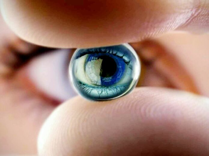
For years, researchers have been working toward restoring even a small semblance of sight for the blind using artificial retinas using a technique called retinal prostheses. However, the images restored following a decade’s worth of studies are still far from being clear.
Until now, that is.
RETINAL PROSTHESES
Retinal prosthesis involves three elements. First, a camera needs to be inserted in the patient’s glasses, followed by an electronic microcircuit, which transforms data taken by the camera into an electric signal. Finally, a matrix of microscopic electrodes are implanted into the eye in contact with the retina. Just like the photoreceptor cells of the retina, the implant converts visual information into electrical signals, which, in turn, are transmitted to the brain via the optic nerve.
In theory, retinal prostheses should help totally blind patients recover visual perceptions in the form of light spots, or phosphenes. But currently, the light signals perceived are not clear enough for a patient to fully recognize a face, read, or move about independently.


NATURAL SIGHT
Scientists from the Institut de Neurosciences de la Timone, AP-HM, CEA-Leti, Institut de la Vision, and Aix-Marseille Universite may have found a way to improve the precision of prosthetic activation.
After comparing the activity of the visual cortex generated by implants against that produced by “natural sight,” the team found that the prosthesis activated the visual cortex of the subjects (i.e. rodents) in the correct position and at ranges comparable to those obtained under natural conditions. However, the extent of the activation was much too great, and its shape was elongated.
In the study published by eLife, the researchers explained that the deformation was due to two separate phenomena observed at the level of the electrode matrix. Firstly, the scientists observed excessive electrical diffusion: the thin layer of liquid situated between the electrode and the retina passively diffused the electrical stimulus to neighboring nerve cells. Secondly, they detected the unwanted activation of retinal fibers situated close to the cells targeted for stimulation.
Using the results of the study, the team was able to improve the properties of the interface between the prosthesis and retina, opening the way towards making promising improvements to retinal prostheses for humans.
This article was provided by Futurism
Best Regards
TBU NEWS


